► Patient Information:
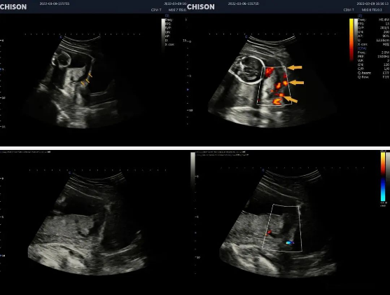
A 27-year-old female with full term pregnancy with a complaint of abdominal pain with massive vaginal bleeding.
► Patient History:
Internal Vaginal Bleeding with intermittent pain treated with medication.
► Scan Impression:
An ultrasound image shows the placenta covering the internal orifice of the cervix and the fluid pocket near the margin of the cervix of about 27*18mm.
Conclusion:
Complete placenta previa with placental abruption.
Ultrasound findings of normal placenta
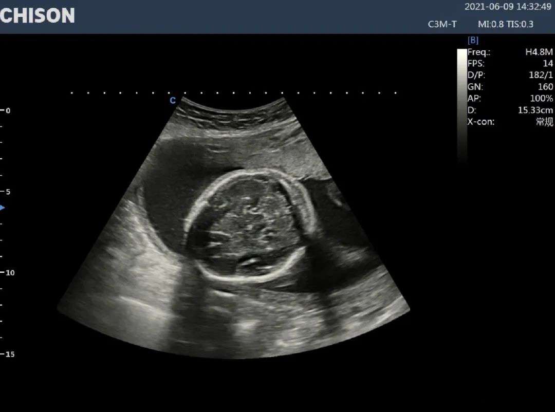
Ultrasound can show the placenta as early as 6 weeks of pregnancy (transvaginal ultrasound) or 10 weeks of pregnancy (transabdominal ultrasound), showing a thin ring, and hypoechoic around the embryo. At the 12th and 13th weeks of pregnancy, a Doppler ultrasound can show the chorionic blood flow. At 14-15weeks of gestation, the placenta is fully developed, which is usually hyperechoic in nature. In the second trimester, the placenta gradually matures and becomes larger, showing a more homogeneous hyperechoic, in which there may be a low echo area with unclear boundaries, which is the placental lake. In late pregnancy, a Doppler ultrasound can show abundant blood flow in the placenta.
Difficulties and differential diagnosis of placental abruption
Placental abruption is one of the obstetrical emergencies that endanger the lives of mothers and infants. The domestic incidence rate is 0.46% - 2.1%. It is a serious complication during pregnancy and a common cause of prenatal bleeding. Untimely treatment can affect the lives of mothers and fetuses.
Ultrasonography is not only an important auxiliary means for the diagnosis of placental abruption but also an important link to improving the maternal and fetal prognosis of placental abruption. It has been widely used in the diagnosis and evaluation of placental abruption. Due to the difference in exfoliation location and area, the performance of placental abruption is ever-changing, placental abruption with typical clinical symptoms, combined with ultrasound examination, is easy to be diagnosed; such as placental abruption with atypical clinical symptoms, it has the characteristics of hidden onset, unobvious clinical manifestations and ever-changing ultrasound images, which is more likely to lead to misdiagnosis and missed diagnosis, bringing greater risk to pregnant women and fetuses. Therefore, atypical placental abruption should be repeatedly examined and carefully screened by ultrasound doctors combined with clinical symptoms and signs.
Ultrasound in Obstetrics and Gynecology
XBit 90 is the flagship product of CHISON's high-end intelligent ultrasound XBit series, which protects women's entire pregnancy. When XBit 90 is used in the examination and diagnosis of placental abruption, the innovative intelligent high-precision imaging will clearly show the changes in the internal structure and shape of the placenta during placental abruption, which will help ultrasound doctors to diagnose timely and accurately and avoid adverse pregnancy outcomes.
Width Enhancement Technology (WET)
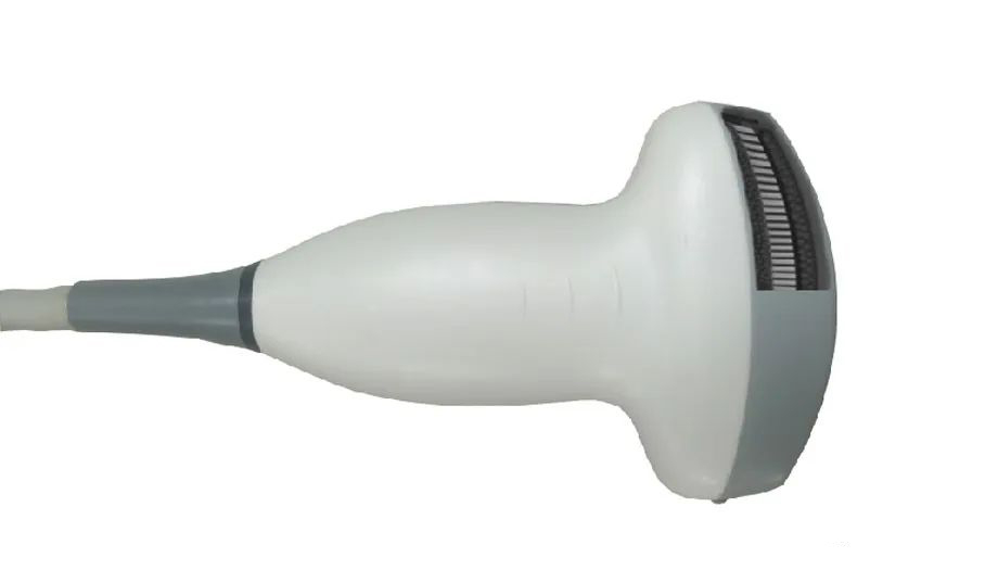
The abdominal wall of some pregnant women is thick, and the examination is relatively difficult, so the probe is required to have sufficient penetration power. XBit 90 Width Enhancement Technology (WET), the probe adopts excellent piezoelectric ceramic composites or piezoelectric single crystal materials, which is made by increasing the gradient of multi-layer matching layer and high-density precision cutting technology, which greatly reduces sound attenuation, effective heat dissipation, broadens the probe frequency band, ensures image resolution and increases good penetration force, calmly dealing with the thicker abdominal wall or front wall placenta.
Microvascular Imaging (MVI)
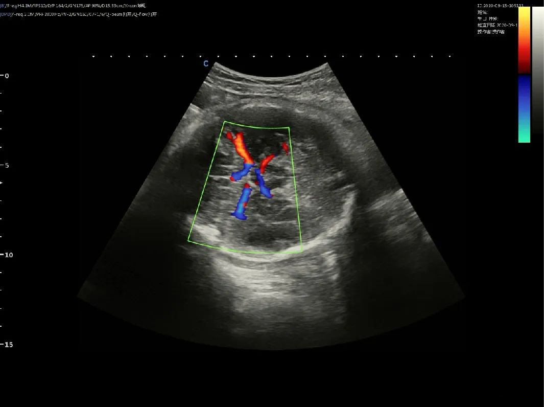
Fetal intracranial flow imaging
Image Optimization Technology (X-ppi)
Because of the ever-changing ultrasound images, atypical placental abruption is easy to be confused with other diseases. The accuracy depends on the clinical experience of ultrasound doctors and the clarity of ultrasound images. XBit 90 Image Optimization Technology (X-ppi), equipped with the NIT platform, quickly acquires every image, finely optimizes each pixel, increases image resolution, improves internal differences of organs or lesions, and high-quality imaging assists in accurate clinical diagnosis.
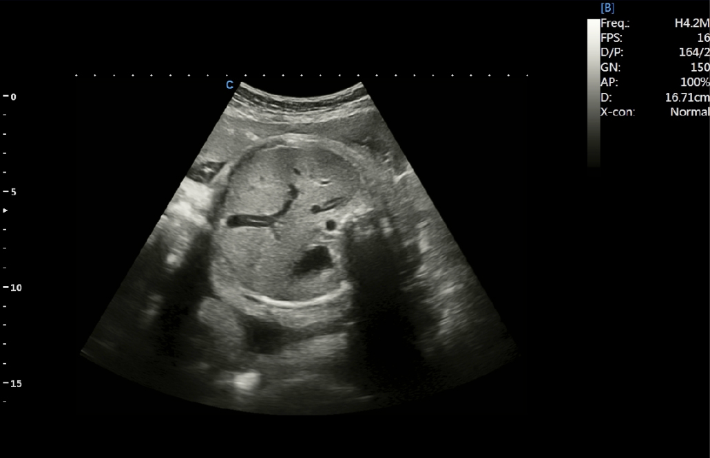
Fetal abdominal section
In the field of obstetrics and gynecology, XBit 90 has a wealth of clinical solutions to meet the needs of health examination and diagnosis. Breast, pre-pregnancy follicle assessment, early pregnancy, and middle and late pregnancy examinations take care of women's entire pregnancy; XBit 90 also pays attention to infants and fetuses, and evaluates their growth and development. CHISON has been making efforts in reproductive health diagnosis and developed a comprehensive detection solution throughout the whole reproductive cycle, protecting the lives and health of women and fetuses.
We are an affordable ultrasound device supplier, please feel free to contact us if you need them.
Contact Us
Copyright © CHISON Medical Technologies Co., Ltd. All Rights Reserved | Privacy and terms of use
Products: Vascular Ultrasound Machine MSK Ultrasound Machine Portable Ultrasound Device Portable Ultrasound Machine for Sale Portable Ultrasound Machine For Pregnancy Handheld Ultrasound For Pregnancy Handheld Veterinary Ultrasound Veterinary Ultrasound Machine Portable Vascular Ultrasound Livestock Ultrasound Machine

CHISON respects your privacy. We use cookies to make our site more personal and enhance your experience. Read our Private Policy to learn more about cookies and how to manage them. You agree to our use of these technologies when you visit our site.

We appreciate your feedback
We sincerely invite you to participate in our survey for helping us to improve our digital market.
*1.How fast does the website load?
*2.Does the products displayed on the website interest you?
*3.How easy is it for you to find the information you need?
4.What information or service do you suggest we can offer?


THANKS
Thank you for sharing your thoughts with us. We’re highly appreciated your every feedback.

Sorry!

THANKS
Thank you for sharing your thoughts with us. We’re highly appreciated your every feedback.